Assessment | Biopsychology | Comparative |Cognitive | Developmental | Language | Individual differences |Personality | Philosophy | Social |
Methods | Statistics |Clinical | Educational | Industrial |Professional items |World psychology |
Biological:Behavioural genetics · Evolutionary psychology · Neuroanatomy · Neurochemistry · Neuroendocrinology ·Neuroscience · Psychoneuroimmunology · Physiological Psychology · Psychopharmacology(Index, Outline)
| Brain: Parietal lobe | ||
|---|---|---|
| ] | ||
| {{{Caption}}} | ||
 |
||
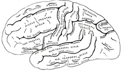
| 左大脳半球の側面、側面から見た。 (頭頂葉は右上にあります。) | ||
| Latin | lobus parietalis | |
| Gray’s | subject #189 822 | |
| Part of | Cerebrum | |
| Components | ||
| Artery | Anterior cerebral Middle cerebral |
|
| Vein | Superior sagittal sinus | |
| BrainInfo/UW | hier-77 | |
| MeSH | A08.186.211.730.885.213.670 | |
頭頂葉は脳の葉です。 それは、後頭葉の上(上)および前頭葉の後ろ(後)に位置する。
頭頂葉は、特に空間感覚とナビゲーションを決定する、異なるモダリティからの感覚情報を統合します。 例えば、それは体性感覚皮質および視覚系の背流を含む。 これにより、頭頂皮質の領域は、視覚的に知覚される物体を身体座標位置にマッピングすることが可能になる。
解剖学
頭頂葉は四つの解剖学的境界によって定義されています:中央溝は頭頂葉を前頭葉から分離し、頭頂-後頭部溝は頭頂葉と後頭部を分離し、側溝(シルビウス裂)は側頭葉からそれを分離する最も外側の境界であり、内側縦裂は二つの半球を分割する。
中心溝のすぐ後部、および頭頂葉の最も前部は、一次体性感覚皮質領域である中心後回(Brodmann領域3)である。 これと後頭頂皮質を分けることは、心後溝である。
後頭頂皮質は、頭頂溝(IP)によって分離された上頭頂小葉(Brodmann領域5+7)と下頭頂小葉(39+40)に細分することができる。 頭頂溝および隣接する回は肢および眼球運動の指導で必要であり、cytoarchitecturalおよび機能相違に基づいて中間(MIP)、側面(唇)、腹側(VIP)、および前方(AIP)区域に更に分けられ
関数
頭頂葉は、身体のさまざまな部分からの感覚情報、数とその関係の知識、およびオブジェクトの操作を統合する上で重要な役割を果た 頭頂葉の一部は視覚空間処理に関与している。 この葉については、大脳の他の三つよりもはるかに少ないことが知られています。
1990年代の様々な研究では、マカクの頭頂皮質の異なる領域が空間の異なる部分を表すことが分かった。
- 外側頭頂内(LIP)には、空間的位置の顕著性を表す網膜局所的にコード化された空間の2次元地形図が含まれています。 それは目の動きを目標とするためにoculomotorシステムによって適切なとき使用することができます。
- 腹側頭頂内(VIP)領域は、多くの感覚(視覚、体性感覚、聴覚、および前庭)からの入力を受け取ります。 触覚受容野を有するニューロンは,頭部中心の基準フレーム内の空間を表現した。 視覚受容野を有する細胞は、頭部中心の基準フレームでも発射するが、おそらく眼中心の座標でも発射する
- 内側頭頂内(MIP)領域ニューロンは、眼中心の座標でリーチターゲットの位置をエンコードする。
- 前頭頂内(AIP)領域には、把持される物体の形状、大きさ、向き、および手自体の操作に応答するニューロンが含まれています。
病理学
Gerstmann症候群は、優性(通常は左)頭頂葉の病変と関連している。 Balintのシンドロームは両側のある損害と関連付けられます。 Hemispatial無視の症候群は、通常、非支配的な半球の注意の大きな赤字と関連している。
このギャラリーに写真を追加
も参照してください
- 脳の葉
- 感覚系障害
- Blakemore&Frith(2005)。 学習脳。 ブラックウェル・パブリッシング所属。 ISBN1-4051-2401-6
- 2.0 2.1 2.2Avillac M,Deneve S,Olivier E,Pouget A,Duhamel JR.(2005)頭頂皮質における視覚的および触覚的位置を表すための参照フレーム。 ナト-ノイローゼ。 8(7):941-9.
- チャンT、ホイヤー HW、ブリテンKH。 (2004)頭部刺激に対する頭頂部VIP神経応答は、頭部中心座標にコードされている。 ニューロン42(6):993-1001.
- Pesaran B,Nelson MJ,Andersen RA. (2006)背側運動前ニューロンは、リーチ計画中に手、目、および目標の相対的な位置をエンコードする。 ニューロン51(1):125-34.
- 5.0 5.1Murata A,Gallese V,Luppino G,Kaseda M,Sakata H.(2000)サルの頭頂部aipのニューロンにおける把持のための物体の形状、大きさ、向きに対する選択性。 J神経生理学83(5):2580. PMID10805659
- Murata A,Gallese V,Kaseda M,Sakata H.(1996)メモリガイド付き手操作に関連する頭頂ニューロン。 J Neurophysiol75(5):2180-6. PMID8734616
終脳(大脳、大脳皮質、大脳半球)-編集
原発性溝/亀裂: medial longitudinal, lateral, central, parietoöccipital, calcarine, cingulate
frontal lobe: precentral gyrus (primary motor cortex, 4), precentral sulcus, superior frontal gyrus (6, 8), middle frontal gyrus (46), inferior frontal gyrus (Broca’s area, 44-pars opercularis, 45-pars triangularis), prefrontal cortex (orbitofrontal cortex, 9, 10, 11, 12, 47)
parietal lobe: postcentral sulcus, postcentral gyrus (1, 2, 3, 43), superior parietal lobule (5), inferior parietal lobule (39-angular gyrus, 40), precuneus (7), intraparietal sulcus
occipital lobe: primary visual cortex (17), cuneus, lingual gyrus, 18, 19 (18 and 19 span whole lobe)
temporal lobe: transverse temporal gyrus (41-42-primary auditory cortex), superior temporal gyrus (38, 22-Wernicke’s area), middle temporal gyrus (21), inferior temporal gyrus (20), fusiform gyrus (36, 37)
limbic lobe/fornicate gyrus: cingulate cortex/cingulate gyrus, anterior cingulate (24, 32, 33), posterior cingulate (23, 31),
isthmus (26, 29, 30), parahippocampal gyrus (piriform cortex, 25, 27, 35), entorhinal cortex (28, 34)
subcortical/insular cortex: rhinencephalon, olfactory bulb, corpus callosum, lateral ventricles, septum pellucidum, ependyma, internal capsule, corona radiata, external capsule
hippocampal formation: dentate gyrus, hippocampus, subiculum
basal ganglia: striatum (caudate nucleus, putamen), lentiform nucleus (putamen, globus pallidus), claustrum, extreme capsule, amygdala, nucleus accumbens
Some categorizations are approximations, and some Brodmann areas span gyri.
This page uses Creative Commons Licensed content from Wikipedia (view authors).